
Introduction:
Suturing is an essential skill that every medical student and surgeon must master. From the selection of sutures, to the selection of different suture methods for different wounds, to the suture techniques under laparoscopy, etc., even every hospital and every doctor has their own suture experience and secrets. Almost no doctor has not been troubled by the "simple" problem of suture during his growth.
The main purpose of suture: to close the wound edge, eliminate dead space, promote healing, and also stop bleeding, rebuild organ structure or plastic surgery;
Suture principles:
1. Strictly abide by aseptic operation.
2. Thorough hemostasis and debridement must be performed before suture.
3. When suturing, the needle should be inserted vertically and removed according to the arc of the needle
4. The suture should be performed in layers according to the anatomical layers of the tissue, leaving no dead space
5. For each layer of suture, the number of groups on both sides of the incision should be equal
6. In order to reduce foreign matter in the incision, the number of sutures should not be too many, generally with a spacing of 0.5~1cm, and try to ensure that the tension of each needle applied to the tissue is equal
First, let's get to know the basic information of sutures:
Model:
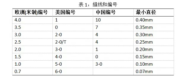
那么,什么情况下用中国编号,什么时候用其它编号呢?在实际工作中,丝线(简称“线”)通常用中国编号,而其它合成缝线使用美国编号。So, when do we use Chinese numbers and when do we use other numbers? In actual work, silk thread (referred to as "thread") is usually numbered in China, while other synthetic sutures use American numbers.
Materials:

Packaging:
There is a lot of useful information on the suture packaging, as shown in the two pictures below:
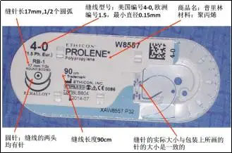
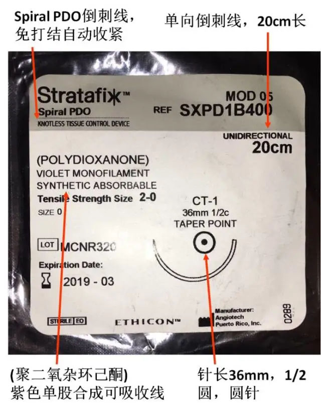
Sutures can be said to be surgical supplies used by surgeons every day. If young doctors can be familiar with the type, material and structure of sutures and can accurately obtain the information on the suture packaging, it will be of great help to the successful completion of the operation.
Suture methods
There are many methods, which can basically be divided into three categories: simple suture, inversion suture and eversion suture. Each category has two types: intermittent and continuous. The choice of suture method is mainly based on the treatment purpose and tissue structure. The following are several commonly used suture methods for understanding
Simple suture method
(1) Simple intermittent suture
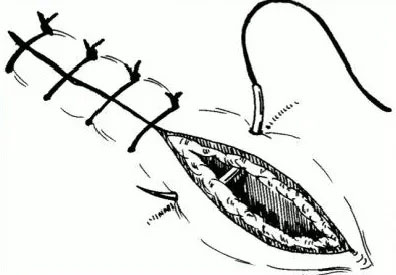

Key points: The most commonly used suturing method, each stitch is tied with a separate knot. It is generally used for suturing various tissues such as skin, subcutaneous tissue, muscle, fascia, peritoneum, etc., especially for suturing infected wounds or wounds with the possibility of infection.
Advantages: Simple and quick operation; firm and reliable suturing. The tension of the incision is shared by each independent knot. After one stitch is removed, it will not affect the entire incision.
Disadvantages: Time-consuming operation, high suture consumption; there is also the possibility of uneven tissue alignment.
(2) Figure 8 suturing
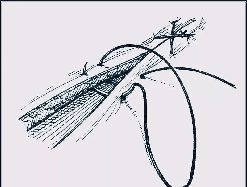

Key points: Also known as double interrupted suture, it is commonly used for suturing the abdominal cavity, white line, subcutaneous tissue, tendons, ligaments, muscles, or suturing and hemostasis of larger blood vessels.
Advantages: Reduce cutting injuries, avoid ischemic necrosis of cut edge tissue, save time, suture firmly, not easy to slip, and can concentrate pressure on the center to stop bleeding.
Disadvantages: The operation is cumbersome.
(3) Simple continuous suture
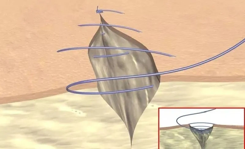
Key points: It is often used for wounds with elasticity and low tension, such as subcutaneous tissue, fascia, pleura, and peritoneum.
Advantages: Tight suture, equal tension, fast and time-saving. The wound edges are tightly aligned and the bleeding is completely stopped.
Disadvantages: If the suture breaks at one place, the entire wound will collapse.
(4) Continuous lock-edge suture
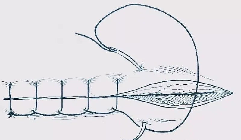

Key points: Also known as blanket suture, the threads are interlaced and fixed each time during the suturing process to form a "locking edge" effect. It is often used for full-thickness suture of the posterior wall during gastrointestinal anastomosis, closure of the gastrointestinal stump, edge suture of the entire free skin graft, suture of the vaginal stump after total hysterectomy, etc.
Advantages: Time-saving operation, good closure and hemostasis effect, more neat and tight suture.
Disadvantages: Relatively time-consuming.
(5) U-shaped imbricate mattress suture
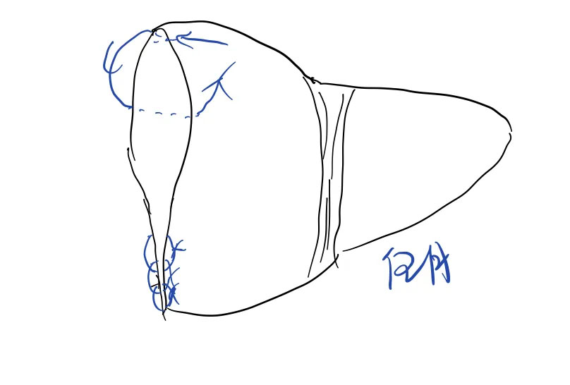
U-shaped imbricate mattress suture: suitable for suturing the cross-section of solid organs such as liver, pancreas or spleen.
Insert the needle from the capsule on one side of the wound edge, penetrate the organ substance to the capsule on the opposite side and then exit the needle. Then insert the needle from the capsule on the same side of the wound edge, penetrate the organ substance to the capsule on the opposite side and then exit the needle. The two ends of the suture are tied on one side of the wound edge. The adjacent two needles overlap. By squeezing the tubular structure of the wound edge, the effect of hemostasis or preventing fluid leakage is achieved.
Eversion suture
After vascular anastomosis, the vascular edge tissue on both sides of the anastomosis is turned outward, while the inner wall of the blood vessel is smooth, with less thread ends left, to avoid thrombosis. It is commonly used for vascular anastomosis, peritoneal suture, tension-relieving suture, etc., and sometimes also used for suturing loose skin to prevent the skin edge from rolling inward and affecting healing.
Specific details are as follows:
(1) Intermittent vertical mattress eversion suture
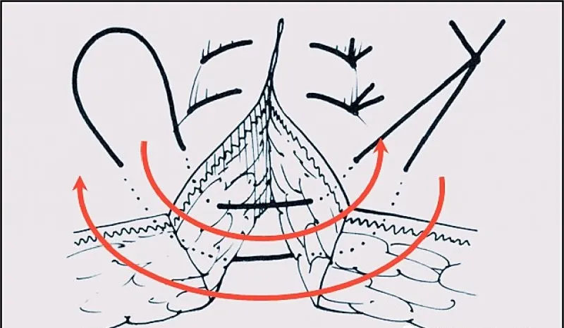

Key points: It is a tension suture, which can be summarized as "far in and far out, near in and near out". The specific method is: insert the needle 5 mm from the incision edge, pass through the epidermis and dermis, cross the incision through the subcutaneous tissue to the opposite side at a symmetrical point 5 mm from the incision edge, then insert the needle from the side where the needle exits 1 to 2 mm from the incision edge, and exit the skin from the opposite side 1 to 2 mm from the incision edge. The plane connected by the four entry and exit points should be perpendicular to the incision, and the skin edges on both sides should be everted.
Applicable: loose skin such as scrotum, axilla, groin, neck, etc.
Advantages: It has strong tensile strength and has little effect on the blood supply of the wound edge.
(2) Intermittent horizontal mattress eversion suture
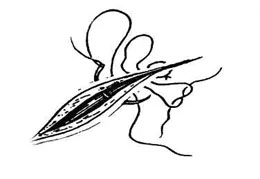
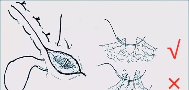

Key points: Insert the needle into the skin 2-3mm away from the incision edge, pass through the epidermis and dermis, cross the incision through the subcutaneous tissue to the corresponding part on the opposite side, then the suture is parallel to the incision and forward about 8mm, then insert the needle again, pass through the skin, cross the incision to the corresponding part on the opposite side, and finally tie a knot with the other end of the suture.
Application: Repair of vascular rupture holes, reinforcement of vascular anastomosis leakage.
Advantages: Fast operation speed, saving sutures, with certain tension resistance conditions, placing a rubber tube on the suture can increase the tension resistance.
Inversion suture
After suturing, the serosal layer is tightly aligned, which is conducive to wound adhesion and healing. After healing, the wound surface is smooth and reduces adhesion with adjacent tissues, preventing wound non-healing or gastrointestinal fluid and urine leakage caused by mucosal eversion.
Details are as follows:
(1) Intermittent vertical mattress inversion suture
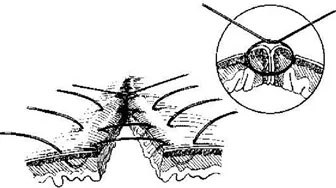

a) Features: The suture passes perpendicular to the incision edge and does not penetrate the intestinal wall mucosa
b) Key points: The needle is inserted into the serosal layer 4-5 mm from the incision edge on one side, and the suture passes between the seromuscular layer and the mucosal layer, and the needle is removed from the ipsilateral serosal layer 2 mm from the incision edge. The needle is inserted across the anastomosis and inserted into the serosal layer 2 mm from the incision edge on the opposite side, and the needle passes between the seromuscular layer and the mucosal layer, and the needle is removed from the serosal layer 4-5 mm from the incision edge. After tying the knot, the intestinal wall of the anastomosis is inverted and buried.
c) Application: The most commonly used inversion suture method for gastrointestinal surgery, which can strengthen the anastomosis and reduce the tension of the anastomosis after full-thickness anastomosis of the gastrointestinal tract.
(2) Continuous full-thickness parallel mattress inversion suture
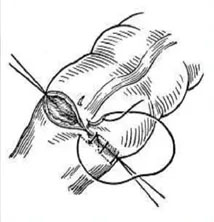
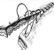
a) Key points: Start with the first stitch to suture the entire thickness of the intestinal wall. That is, insert the needle from one side of the serosa and pass through the entire thickness, then insert the needle from the opposite side of the mucosa and exit from the serosa. After tying a knot, insert the needle from the serosa 3-4 mm away from the knot and pass through the entire thickness of the intestinal wall. Then insert the needle from the mucosa of the ipsilateral intestinal wall and exit from the serosa. When the suture reaches the contralateral intestinal wall, insert and exit the needle in the same way, tighten the suture to turn the incision edge inward. Note that the ipsilateral entry and exit points are 2 mm away from the incision edge, and the line connecting the entry and exit points should be parallel to the incision edge.
b) Application: Mostly used for full-thickness suture of the anterior gastrointestinal wall and suture of the uterine wall.
(3) Continuous horizontal mattress-style seromuscular layer inversion suture

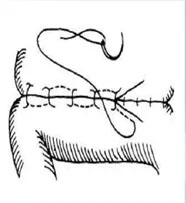
a) Key points: The suturing method is similar to the Connell suture, but the suture layer is different. This method only passes through the seromuscular layer instead of the entire layer. See the identification diagram for details.
b) Application: It can be used for anastomosis of the seromuscular layer of the anterior and posterior walls of the gastrointestinal tract.
The suture does not penetrate the mucosa, but passes between the seromuscular layer and the mucosal layer, turning the tissue at the wound edge inward, keeping the outer surface smooth and well aligned, and reducing adhesion. It is often used for anastomosis of the gastrointestinal tract and bladder.
(4) Purse-string suture
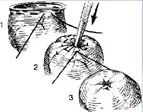
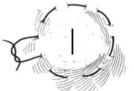
a) Key points: It is an inversion suture within a small range. The seromuscular layer is sutured continuously in a circle for one week at the intended embedding site. After ligation, the center is inverted and embedded, and the surface is smooth, which is conducive to healing and reduces adhesion.
b) Application: It is often used for embedding the appendix stump, fixing the ostomy tube of the hollow organ, sealing the small wound or puncture needle eye of the gastrointestinal wall, fixing the ostomy tube of the hollow organ, etc.
Tension-reducing suture
When the tissue tension at the suture site is large and the systemic condition is poor, this method can be used to prevent the incision from splitting. It is mainly used for tension reduction of the abdominal wall incision.
Use thicker silk thread for sutures, insert the needle 2~2.5 cm away from the wound edge to ensure the accuracy of the layer, and the distance between sutures is 3~4 cm. Make it bear more incision tension. Before ligation, pass the suture through a rubber tube or gauze pillow to prevent the skin from being cut. Do not tie too tightly during ligation to avoid affecting blood circulation.
Suture tension strength: tension-reducing suture > 8-shaped suture, U-shaped suture > interrupted suture.
The above surgical suture methods are only used as a guide for teaching and reference, and may be slightly different from actual clinical applications. Each surgeon has different suture habits and suture methods, but as medical students and young doctors, they still need to master the most basic and classic things.